Scientists have achieved a revolutionary feat by effectively using ultrasonic waves to probe the inner workings of a human brain. This groundbreaking study represents the first instance of real-time monitoring and recording of brain activity while an individual is performing tasks outside of a standard medical setting, like playing a video game.
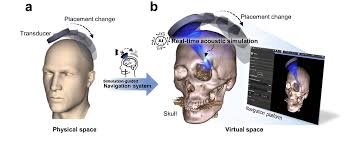
The researchers’ novel strategy included inserting a certain substance into the subject’s skull. Because of the material’s “acoustically transparent” window, ultrasonic vibrations were able to enter the brain. Once within, these waves interacted with various tissues, returning to an ultrasonic probe attached to a scanner after bouncing off limits. Then, using data processing techniques, a precise picture of brain activity was created, similar to the way ultrasound scans show images of developing fetuses.
The primary goal of the research was to track blood volume variations in two distinct brain regions—the motor cortex and the posterior parietal cortex. These areas are critical for a number of cognitive processes and for coordinating movement.This method’s primary tenet is the indirect monitoring of brain cell activity via variations in blood volume. Neurons demand more oxygen and nutrients from blood arteries when they become more active. Researchers can learn more about the dynamic activity of brain cells throughout various tasks and activities by evaluating changes in blood volume.
This ground-breaking study applies earlier findings from studies on nonhuman primates to a human participant. Using ultrasound imaging, the team was able to see and examine the complex neuronal activity that was taking place in the person’s brain while he performed a variety of tasks, such as strumming a guitar or playing a game of connect-the-dots.
The results of this investigation, which were released on May 29 in the journal Science Translational Medicine, mark a substantial advancement in our knowledge of brain activity and function. The study used a multimodal strategy that combined cutting-edge imaging techniques with novel technologies. The subject’s skull had to have a particular substance implanted as the initial stage. This substance made it easier for ultrasonic waves to enter the brain, creating a direct line of sight for imaging and tracking brain activity.
The ultrasonic waves interacted with many tissues, such as neurons, blood vessels, and other structural elements, once they had entered the brain. These waves caused boundaries between tissues to reflect, producing useful data that an ultrasound probe attached to a scanning equipment was able to record.
By capturing real-time variations in blood volume inside specific brain regions, the scanning procedure offered a dynamic perspective on neuronal activity. This method gave researchers insight into the complex systems underpinning cognition and movement by allowing them to watch how brain cells behaved and cooperated during different tasks.
Furthermore, blood volume variations can be tracked to provide a more complex picture of brain activity. A better understanding of neurovascular coupling—the complex interaction between neuronal signaling and vascular responses—is made possible by the link between brain activity and blood flow.
The research has ramifications that go beyond academic curiosity. It has potential applications in the fields of healthcare, neurorehabilitation, and cognitive enhancement. The capacity to see brain activity in real-time without invasive methods is extremely valuable for the diagnosis of neurological conditions, evaluation of cognitive abilities, and development of individualized treatment plans.
Ultrasound imaging has the potential to transform neuroimaging protocols in clinical settings by providing a more affordable and accessible substitute for conventional techniques. Because ultrasound equipment is portable, patients with neurological disorders can be monitored at the bedside, allowing for prompt interventions and customized treatment plans.
Moreover, therapies targeted at improving motor and cognitive abilities can benefit from the knowledge gathered from researching brain activity during particular tasks. Through comprehending how brain circuits adjust and react to distinct stimuli, scientists may create focused interventions for improving neuroplasticity, cognitive training, and rehabilitation.
One interesting area of neurotechnology is the incorporation of ultrasound imaging into brain-computer interfaces, or BCIs. Direct brain-to-external device communication is made possible by BCIs, which may find use in neuromodulation treatments, prosthetics, and assistive technology.BCIs may read brain signals more accurately and responsively by utilizing real-time neural monitoring capabilities, which enhances the effectiveness and user-friendliness of these technologies. A new era of neuroengineering is being ushered in by the synergy between ultrasound imaging and BCIs, where precise neural control and feedback open the door to creative solutions in healthcare and human augmentation.
Furthermore, learning about brain activity in various settings advances our comprehension of human consciousness, behavior, and thought. This information drives advances in neuroscience, encouraging multidisciplinary cooperation and breaking through conventional barriers in research.


