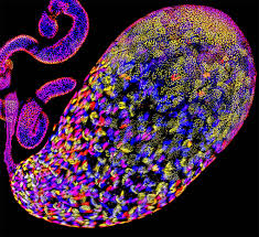
How the human body detects heat has long been a biological puzzle, but scientists at Northwestern University have now uncovered new details about a key molecular heat sensor. Their study reveals how a protein called TRPM3 helps nerve cells detect rising temperatures — and challenges previous assumptions about how heat is sensed at the cellular level.
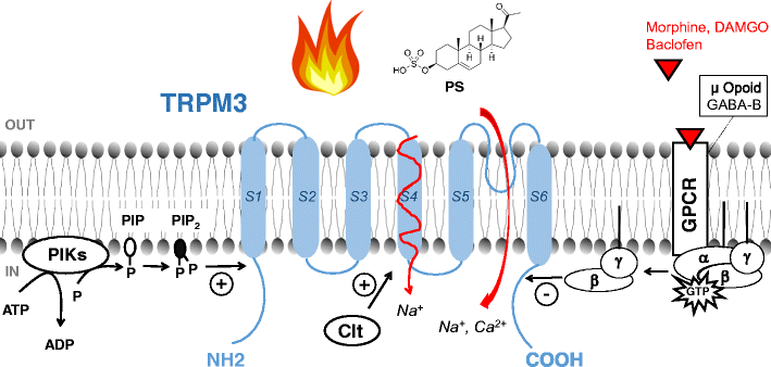
TRPM3 is a channel protein that sits in the membrane of sensory nerve cells. When activated, it opens like a gate to allow charged particles (ions) to move into the cell. This ion flow generates electrical impulses that the nervous system interprets as heat or pain. Previously, scientists believed that TRPM3 detected heat through the portion of the protein embedded in the cell membrane. However, the new study shows that the heat-sensitive region actually lies inside the cell, on the inner section of the protein.
This discovery not only reveals a new mechanism of temperature detection but also explains how the body can distinguish between mild warmth and dangerously high heat. Because TRPM3 is also linked to pain perception, inflammation, and neurological disorders such as epilepsy, the findings could accelerate the development of new non-opioid pain therapies.
To study TRPM3, the Northwestern team used cryo-electron microscopy (cryo-EM) to capture high-resolution 3D images of the protein in different states. They induced an “active” heat-like state using a chemical activator and froze the protein in action. They also mapped its structure in an “inactive” state using an epilepsy drug known to block TRPM3. Comparing the two states revealed how parts of the protein shift during activation.
Their results demonstrate that both heat and chemicals trigger similar internal structural movements in TRPM3, offering a detailed view of how temperature activates the channel. The study will be published October 24 in Nature Structural & Molecular Biology.