Scientists Develop Cellular Models to Unlock Embryo Development Mysteries
UC Santa Cruz Researchers Create Programmable Embryo-Like Structures Using CRISPR
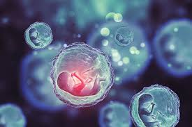
The early stages of embryo development remain one of the greatest scientific puzzles. Once a sperm fertilizes an egg, a complex process begins, transforming a single cell into a fully developed organism. For many species, this transformation occurs within the protective confines of the uterus, making direct observation and study difficult. As a result, researchers have long struggled to understand developmental errors, risk factors, and environmental influences that could prevent successful embryo formation.
Now, scientists at the University of California, Santa Cruz (UCSC) have made a breakthrough by engineering cellular models of embryos without using actual embryos. By leveraging CRISPR-based technology, they have been able to mimic the first few days of development, allowing for detailed studies of gene function in early embryonic formation. Their findings were recently published in Cell Stem Cell, a leading journal in stem cell research.
Innovating Embryo Research Without Embryos
Assistant Professor Ali Shariati, a biomolecular engineering expert and senior author of the study, emphasized the importance of recreating biological phenomena in a controlled laboratory environment.
“As scientists, we strive to replicate and modify natural processes to enable studies that would otherwise be impractical,” said Shariati. “Our goal is to understand how cells self-organize into embryo-like structures and identify factors that may disrupt development.”
Rather than working with actual embryos, UCSC researchers developed lab-grown structures known as embryoids. These are clusters of stem cells that self-organize to mimic certain aspects of early embryonic development, providing a powerful tool to explore genetic influences without ethical concerns.
Breakthrough in Stem Cell Engineering
The research, led by postdoctoral scholar Gerrald Lodewijk and alumna Sayaka Kozuki, utilized mouse stem cells commonly cultivated in laboratories. The team employed an advanced version of CRISPR technology known as an epigenome editor, which does not alter DNA sequences but modifies how genes are expressed. By targeting key genomic regions involved in embryo formation, they were able to control gene activation and guide cells into developing as they would in a natural embryo.
“Stem cells act as a blank canvas,” Lodewijk explained. “By using CRISPR tools, we can guide them to differentiate into various cell types that contribute to early embryonic development.”
This method proved to be more natural than previous chemical-based approaches, as it allowed different cell types to develop together in a process resembling real embryonic growth. Remarkably, 80% of the stem cells successfully organized into structures that reflected key features of early embryos.
Observing Collective Cellular Behavior
One of the most fascinating discoveries was how the cells behaved collectively, moving and interacting in ways similar to naturally forming embryos. Researchers observed that some cells exhibited coordinated rotational migration, resembling patterns seen in flocks of birds or schools of fish.
“The way these cells move and organize is astonishing,” Shariati noted. “They require minimal external input—it’s as if they already have an inherent blueprint, and we merely provide guidance.”
Programmable Embryo Models for Genetic Research
A major advantage of these models is their programmability. Researchers can manipulate gene activation at various stages, testing how specific genetic changes impact development. This approach provides a more comprehensive view of early embryogenesis and could help scientists uncover causes of developmental disorders and genetic mutations.
“Our models offer a more complete representation of early development,” said Lodewijk. “By precisely controlling gene activation, we can investigate the effects of multiple genetic factors on embryonic formation.”
In a demonstration of the technology’s potential, the team identified how certain tissue formations could be either promoted or inhibited based on gene modifications. Their method could be applied across various species, enabling embryo studies without the need to work with actual embryos.
Implications for Fertility and Reproductive Health
This research could have far-reaching implications, particularly in understanding reproductive challenges in humans. Among mammals, human embryos often struggle with implantation and early structural organization, leading to higher rates of developmental failure.
By identifying critical factors that contribute to these early-stage failures, scientists hope to improve fertility treatments and reproductive health outcomes. The UCSC team’s work could pave the way for breakthroughs in fertility research, offering hope to those facing difficulties in conception.
With their innovative approach, UCSC researchers have provided a new lens through which scientists can study early development, shedding light on one of biology’s most intriguing and elusive processes.